
A team from Lawrence Livermore National Laboratory led by Michael Thelen, in collaboration with researchers from Lawrence Berkeley National Lab and the National Renewable Energy Laboratory, has examined the chemical and structural organization of the plant cell wall in Zinnia elegans tracheary elements (TEs). Tracheary elements are elongated cells in the xylem of vascular plants that serve in the transport of water and mineral salts.
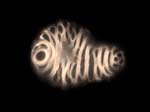
A xylem cell with fluorescent lignocellulose bands as the major feature.
Using three different microscopy methods (AFM, synchrotron radiation-based FTIR
spectromicroscopy, and fluorescence microscopy using a cellulose-specific CBM) in
conjunction with chemical extraction of wall components, the team was able to
visualize single cells in detail down to the nanoscale, cellular substructures, fine-scale organization of the cell wall, and the chemical composition of these cells, indicating that they contain an abundance of lignocellulose.
The Zinnia TE culture system proved ideal for observing the structure and chemical composition of the cell wall because it comprises a single homogeneous cell type, representing a simpler system compared to plant tissues, which may contain multiple cell types.
—Lacayo et al.
The leaves of Zinnia seedlings provide a rich source of single cells that are dark green with chloroplasts and can be cultured in liquid for several days at a time. During the culturing process, the cells change in shape to resemble the tube-like cells that carry water from roots to leaves. Known as xylem, these cells hold the bulk of cellulose and lignin in plants, which are both major targets of recent biofuel research.
The basic idea is that cellulose is a polymer of sugars, which if released by enzymes, can be converted into alcohols and other chemicals used in alternative fuel production. But for this to happen efficiently, we need to find ways to see how this is proceeding at several spatial scales.
—Michael Thelen
The polymers, collectively called lignocellulose, are very insoluble, resistant to common chemicals and mechanical breakage, and are a superior substance for providing strength and structure to plants.
Source: Lacayo et al.
The detailed three-dimensional molecular cell wall structure of plants remains poorly understood.
The capability to image plant cell surfaces at the nanometer scale, together
with the corresponding chemical composition, could significantly enhance our
understanding of cell wall molecular architecture. A high resolution structural model is crucial for the successful implementation of new approaches for conversion of biomass to liquid fuels.—Alex Malkin, a member of the LLNL team who is an expert in atomic force microscopy.
To make fuels from plant biomass requires a thorough understanding of the organization of cell walls before determining the best methods for cell wall deconstruction into its components. Catherine Lacayo, a postdoctoral scientist working with Thelen and Malkin, has taken the first steps toward a comprehensive approach.
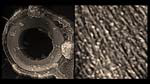
Image showing a substructure of the cell wall (ring), and the detailed organization of lignocellulose in the cell wall.
She came up with techniques that reveal the inner structure of cell walls in these single xylem cells, which represent about 70% of the cellulose in plants that can be used in fuel processing.
The research is supported by the Department of Energy Genome Sciences Program through the Office of Biological and Environmental Research, and the DOE’s BioEnergy Research Centers in Emeryville and Oak Ridge. It will appear in the September issue of Plant Physiology.
Publication:
Catherine I. Lacayo, Alexander J. Malkin, Hoi-Ying N. Holman, Liang Chen, Shi-You Ding, Mona S. Hwang and Michael P. Thelen (2010) Imaging Cell Wall Architecture in Single Zinnia elegans Tracheary Elements. Plant Physiologydoi: 10.1104/pp.110.155242